1
Spondylolysis and Spondylolisthesis
![SPONDYLOLYSIS – shutterstock_1835855416 [Converted]](https://gymnasticsmedicinemens.org/wp-content/uploads/2023/05/SPONDYLOLYSIS-shutterstock_1835855416-Converted-scaled.jpg)
![SPONDY – PHOTO – shutterstock_1360119188 [Converted]](https://gymnasticsmedicinemens.org/wp-content/uploads/2023/05/SPONDY-PHOTO-shutterstock_1360119188-Converted.jpg)
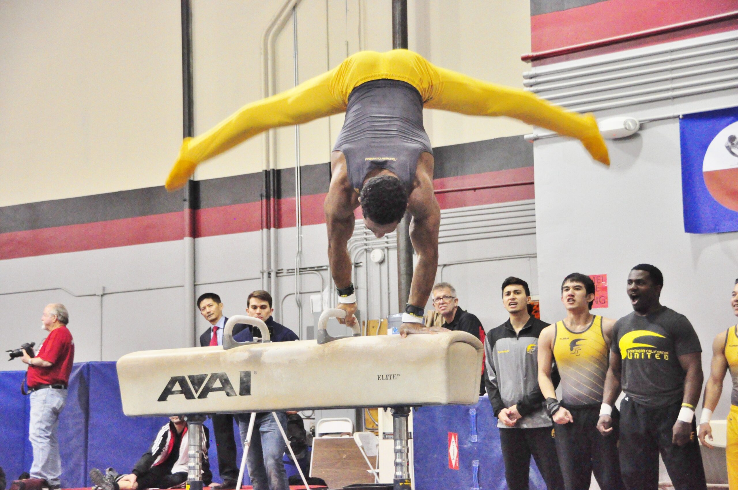
Epidemiology: Typically, this occurs in gymnasts who do repetitive hyperextension (arching) of spine.
Mechanism of Injury/Description: Repetitive hyperextension (arching) and twisting of the lumbar spine causes a fracture or stress fracture at the pars interarticularis (location in the lumbar spine).
Spondylolisthesis: a bilateral Spondylolysis which causes a slipping of the vertebral body.
Signs/Symptoms: Gymnasts typically have low back pain with extension (arching) which has typically been ongoing for a few weeks or months. Sometimes twisting and impact may also cause low back pain.
Diagnosis: On physical exam, gymnasts may be tender to palpation (touch) on the lumbar spinous processes, have pain with extension (arching), and have a positive stork test (pain with extending back on one leg). An x-ray sometimes shows the fracture (but not always and one study by Atsushi Kobayashi et. al. found that 48% were missed on x-ray but seen on MRI), however, your medical provider may recommend an MRI or CT.
Treatment: Your medical provider will likely recommend a period of rest from gymnastics and physical therapy. Sometimes a brace may also be used.
Prevention: To prevent this injury improve core strength and stability, increase shoulder forward flexion, hip extension, and work on posture and proper technique.
Gymnastics Medical Provider PEARLS: Consider ordering a 3T Lumbar Spine MRI to obtain more detail. Not all spondylolysis heal but they can become symptom free and return to the sport with proper medical clearance.
Gymnast, Parent, and Coach PEARLS: To avoid this injury, focus on:
- Limiting arching in the lumbar spine when possible (ex: back walkovers, front walkovers).
- Performing splits on the floor with square hips in all ages.
- Checking to see if your gymnast can lift his/her arms by their ears without moving their body (except arms) and avoid an arch of the lumbar spine.
- Be sure your gymnast is not “hinging” from her lumbar spine with gymnastics skills.
2
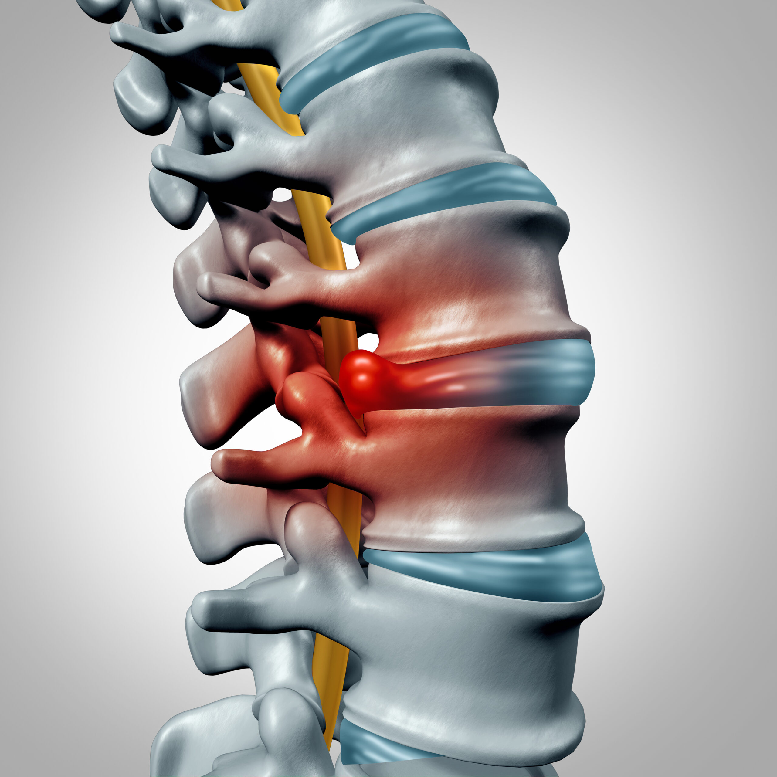
Discogenic Back Pain
Epidemiology: Disc injuries typically occur in gymnasts in their teens and older. There have been some studies that show 11% of children have disc-related pathology compared with 48% of adults.
Mechanism of Injury/Description: Discs of the spine are the “squishy” areas between the vertebrae (bones). They help to absorb impact/forces. They can be injured from a one-time injury or due to repeated impacts. Disc injuries can typically occur from repetitive flexion (touching your toes/ pike/bending forward) or an acute hard/unexpected landing or flexion (touching your toes/ pike/bending forward).
Signs/Symptoms: Gymnasts may have pain with forward flexion (touching your toes/ pike/bending forward) or have nerve pain that causes numbness or tingling into/down your legs.
Diagnosis: Discogenic back pain is typically diagnosed by a physical exam including pain with forward flexion. Your Medical Provider may get imaging studies (ex: x-ray or MRI) of your back to confirm the diagnosis.
Treatment: Treatment may include a brace, rest from impact on the spine, and physical therapy.
Prevention: To prevent this injury focus on core strength, posture training, proper landing mechanics, and proper technique/movement.
Gymnastics Medical Provider PEARLS: Providers should note that disc injuries may be seen on MRIs and could be incidental findings and so it is advised to correlate your physical exam with your imaging. “Traditionally” flexion based back pain is “disc related”.
Gymnast, Parent, and Coach PEARLS: Do not ignore neurologic signs and symptoms this means numbness or tingling down your leg (or legs) or changes to bowel bladder movements. Immediately seek medical attention if your gymnast has these signs/symptoms.
3
Gymnast Back/ Spondylogenic
Mechanical/Functional
Back Pain
Epidemiology: Any age gymnast may have mechanical or functional back pain. This is a diagnosis of exclusion (meaning all other causes were ruled out via physical exam and imaging).
Mechanism of Injury/Description: This diagnosis typically occurs due to repetitive hyperextension (arching), poor posture (rounded shoulders), decreased shoulder forward flexion, and tight hip flexors all lead to increased pressure and loading of the spine causing this overuse injury.
Signs/Symptoms: Gymnasts may have pain with extension (arching), and sometimes pain with flexion (bending forward). Pain is typically non-specific and not in a “pinpoint area” but typically in the low back region. This pain typically has been ongoing for a few weeks or months.
Diagnosis: This is a diagnosis of exclusion-meaning your Medical Provider has ruled out all other causes/diagnoses. Your Medical Provider may diagnose you based on physical exam and imaging (ex: x-rays, MRI, CT scan, etc…).
Treatment: Your Medical Provider may recommend a brace, activity modification in the gym, and physical therapy.
Prevention: You can prevent mechanical back pain by working on core strength/stability, posture, improving shoulder and hip flexibility, and by using proper technique with arching skills.
Gymnastics Medical Provider PEARLS: Consider ordering a 3T Lumbar Spine MRI to obtain more detail. Sometimes on MRI facet inflammation may be seen. Also consider Baastrup’s syndrome if spinous processes are inflamed on MRI or tender on exam.
Gymnast, Parent, and Coach PEARLS: Do not ignore back pain. Back pain treated early is easier to get rid of than back pain which has been there for months or years.
4
Vertebral Body Fracture
Epidemiology: A spine fracture can occur in any gymnast who is performing high risk skills.
Mechanism of Injury/Description: A vertebral body fracture usually occurs from an acute fall in a hyperflexed position or an unexpected downward loading/force.
Signs/Symptoms: Gymnasts will have immediate back pain that is worse with movement and impact.
Diagnosis: Your Medical Provider may suspect a fracture based on the history and exam. An x-ray will show the fracture.
Treatment: Treatment for a vertebral body fracture includes a brace, rest from impact/pounding, and physical therapy.
Prevention: Proper landing techniques and using safe mats can help to prevent a fracture.
Gymnastics Medical Provider PEARLS: Any time there is an acute fall/injury an x-ray is needed to assess the patient further. The gymnast may have a video of the injury or can describe the mechanism which can help you make the diagnosis and plan. Also ask the gymnast/parent/guardian if there is an emergency action plan in place at their gymnastics gym.
Gymnast, Parent, and Coach PEARLS: If a gymnast has an unexpected fall and acute pain do not move the gymnast. Ask someone to call 911 and have the gymnast transported by EMS immediately.
5
Cervical Spine Injury
Epidemiology: Can occur at any age.
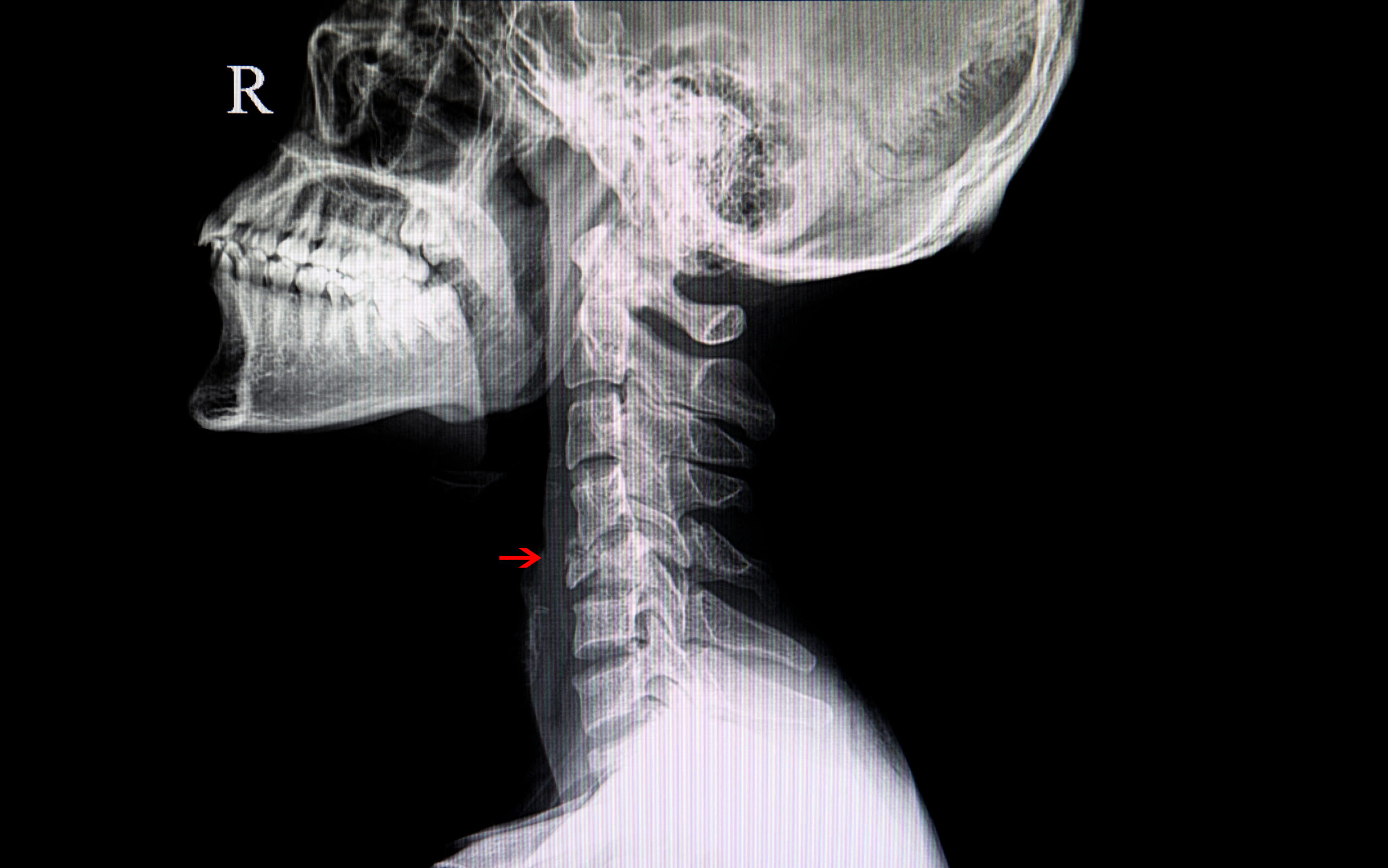
Mechanism of Injury/Description: A cervical spine injury is the result of a direct blow or injury to the neck.
Signs/Symptoms: The gymnast will have acute pain in their neck (cervical spine) and may have numbness or tingling. **If there is any loss of sensation/feeling/numbness, 9-1-1 should be called immediately and as long as the gymnast is in a safe position/area the gymnast should not be moved until EMS or a medical provider has arrived.
Diagnosis: If a cervical spine injury is suspected, and a trained medical provider is available stabilize the head and neck immediately, then log roll (or check your States regulations on emergency training/protocols), so the athlete is prone; finally, depending on your State’s protocols, and if allowed/trained use a spine board, and transport the gymnast to the local emergency department. Once transported, further imaging may be ordered (ex: x-ray, MRI, CT scan).
Treatment: Transportation to your local emergency department.
Prevention: To prevent cervical spine injuries practice proper landing techniques, proper matting and spotting, and neck strengthening.
Gymnastics Medical Provider PEARLS: Know your States individual laws on spine boarding. Talk with gymnastics facilities about practicing spine boarding and cervical spine injury extraction from the foam pit.
Gymnast, Parent, and Coach PEARLS: Ask your coaches/gymnastics facility if they have an Emergency Action Plan (EAP). This plan should be in place for all gymnastic facilities and competition venues.
6
Sacroiliitis
Epidemiology: Sacroiliitis can occur at any age.
Mechanism of Injury/Description: Sacroiliitis is due to inflammation of the sacroiliac joints, or the joint where the low back and pelvis meet. This can be on one side (unilateral), or on both sides (bilateral).
Signs/Symptoms: The gymnast will have pain in the low back/pelvis/buttocks region, and sometimes the pain will radiate down into one or both legs. There can also be pain with prolonged sitting or standing.
Diagnosis: Sacroiliitis is suspected on physical exam when there is pain to palpation/touch on the sacroiliac joints with a positive FABER test, positive Gaenslen test, and positive Sacral distraction test. An x-ray may show an irregularity at the sacroiliac joint. Your Medical Provider may order an MRI to confirm the diagnosis.
Treatment: The gymnast should rest from impact/pounding/force on the sacroiliac joints and attend physical therapy for treatment of sacroiliitis.
Prevention: Teaching gymnasts proper landing, and improving core strength and hip/shoulder flexibility can help to prevent sacroiliitis.
Gymnastics Medical Provider PEARLS: This can be a difficult diagnosis to make because patients may start by complaining about nonspecific low back pain. Physical exam is key to locating the pain on the SI joint. An injection directly in or near the joint (using ultrasound guidance to confirm the location) can be helpful if other conservative treatment has failed.
Gymnast, Parent, and Coach PEARLS: Do not ignore sacrum pain. Sacrum/low back pain treated early is easier to get rid of than sacrum/low back pain which has been there for months or years.
7
Kyphosis
Epidemiology: Kyphosis may occur at any age but is usually seen in adolescents during rapid periods of growth.
Mechanism of Injury/Description: There is no specific cause of kyphosis but sometimes it runs in families or can be due to poor posture.
Signs/Symptoms: Gymnasts with kyphosis have a rounding of the thoracic (mid-back). They may or may not have pain.
Diagnosis: Your Medical Provider will do a physical exam to look for a rounded thoracic spine. An x-ray will show a curve greater than 50 degrees.
Treatment: Your Medical Provider will recommend physical therapy and possibly bracing, depending on the degree of the curve.
Prevention: Working on posture, core strength, and hip/shoulder flexibility can decrease your risk of kyphosis.
Gymnastics Medical Provider PEARLS: Early detection is key and starting physical therapy early can be very helpful.
Gymnast, Parent, and Coach PEARLS: Parents and Coaches if you notice your gymnast is developing poor posture (and having difficulties correcting it) seek medical attention from an Orthopedist or Sports Medicine Provider.
8
Scheuermann’s Disease
Epidemiology: Scheuermann’s Disease typically occurs in late childhood or adolescent athletes.
Mechanism of Injury/Description: Exact cause is unknown but may be related to abnormal bone growth and seems to run in families.
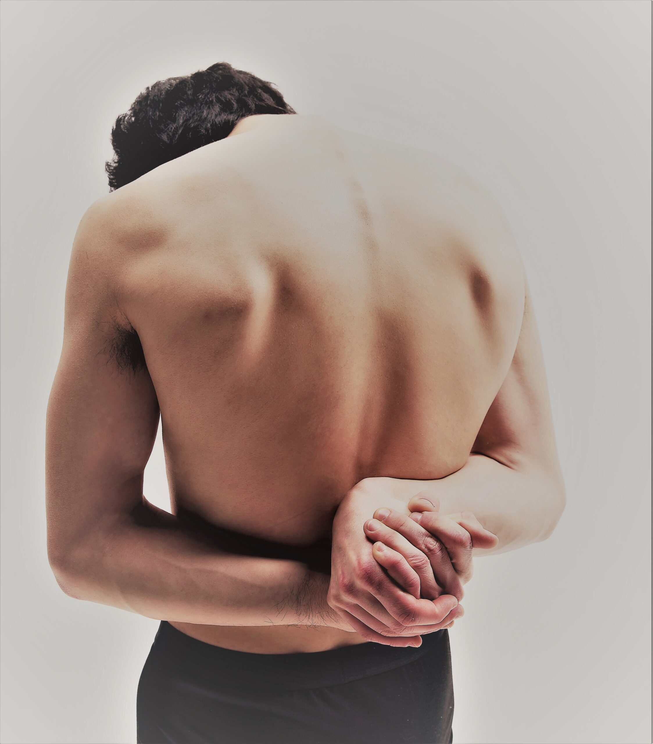
Signs/Symptoms: Gymnasts will have a rounded back when standing and when bending forward. They may have pain in the middle back. In severe cases this may even cause trouble breathing.
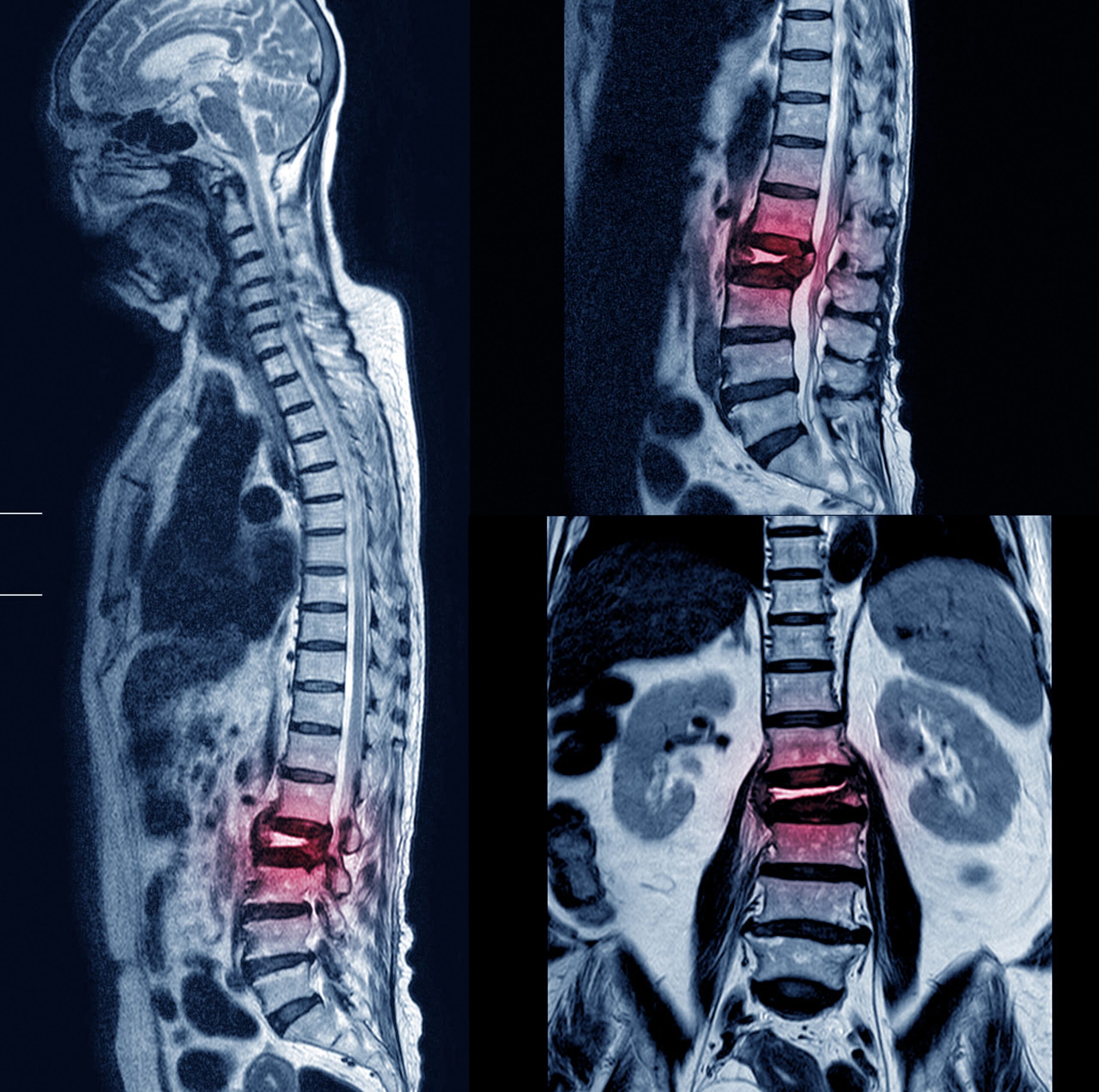
Diagnosis: Your Medical Provider will do a physical exam to look at your posture in the mid-back/thoracic spine (which will be rigid and a “roundback”). X-rays will show wedging of 5 degrees of 3 or more vertebrae, an angle greater than 50 degrees in the thoracic spine (kyphosis) and Schmorl’s nodes, which are irregularities of the vertebral bodies. Occasionally an MRI is performed.
Treatment: Your Medical Provider may recommend a Milwaukee brace or physical therapy.
Prevention: There is no specific prevention, but having a strong core, good posture, proper technique, and limiting impacts can help decrease your risk for Scheuermann’s Disease.
Gymnastics Medical Provider PEARLS: Consider ordering an MRI to investigate other abnormalities in the spinal cord.
Gymnast, Parent, and Coach PEARLS: Parents and Coaches if you notice your gymnast is developing poor posture (and having difficulties correcting it) seek medical attention from an Orthopedist or Sports Medicine Provider.
9
Type II Scheuermann’s Disease
Atypical/Lumbar Scheuermann’s Disease
Epidemiology: Atypical Scheuermann’s occurs in late childhood/adolescents.
Mechanism of Injury/Description: The exact cause is unknown but it seems to run in families. This is similar to Scheuermann’s Disease only this is in the lumbar spine.
Signs/Symptoms: Gymnasts will present with a rounded back and mid or low back pain, especially with forward flexion.
Diagnosis: On physical exam, your Medical Provider may note kyphosis, or rounded back, on forward flexion (bending forward/touching your toes). X-rays may show abnormalities of the vertebral bodies and a straight spine (typically there is no wedging as compared to Scheuermann’s Disease), and you may have Schmorl’s nodes, which are irregularities of the vertebral bodies. Occasionally, an MRI is needed.
Treatment: Your Medical Provider may recommend physical therapy or a lordotic brace with 15° of lordosis.
Prevention: There is no specific prevention, but having a strong core, good posture, proper technique, and limiting impacts can help decrease your risk for Scheuermann’s.
Gymnastics Medical Provider PEARLS: Consider ordering an MRI to investigate other abnormalities in the spinal cord.
Gymnast, Parent, and Coach PEARLS: Parents and Coaches if you notice your gymnast is developing poor posture (and having difficulties correcting it) seek medical attention from an Orthopedist or Sports Medicine Provider.
![SI joint pain – shutterstock_1336785158 [Converted]](https://gymnasticsmedicinemens.org/wp-content/uploads/2023/05/SI-joint-pain-shutterstock_1336785158-Converted.jpg)
![shuermans disease – shutterstock_2159809523 [Converted]](https://gymnasticsmedicinemens.org/wp-content/uploads/2023/05/shuermans-disease-shutterstock_2159809523-Converted-scaled.jpg)